 极端血液稀释对犬脑血流自身调节,脑电图和脑氧代谢率的影响
# 极端血液稀释对犬脑血流自身调节,脑电图和脑氧代谢率的影响
The Effects of Extreme Hemodilutions on the Autoregulation of Cerebral Blood Flow, Electroencephalogram and Cerebral Metabolic Rate of Oxygen in the Dog
Maruyama M, Shimoji K, Ichikawa T, Hashiba M, Naito E. The effects of extreme hemodilutions on the autoregulation of cerebral blood flow, electroencephalogram and cerebral metabolic rate of oxygen in the dog. Stroke. 1985;16(4):675-679. doi:10.1161/01.str.16.4.675
DeepL翻译 + 人工校对# 摘要
SUMMARY The effects of profound (hematocrit value, Ht 20%) and extreme (Ht 5%) hemodilutions on the relationship between the mean arterial pressure (MAP) and the cerebral blood flow (CBF) were studied in pentobarbital-anesthetized dogs. A regression line was found between the CBF and Ht values during normotensive hemodilution (MAP 100 torr): CBF(ml/100g·min) = - 98.9 log Ht (%) + 195.5 (p < 0.001). The CBF was increased by hemodilution, but the range of its autoregulation was narrowed, suggesting a progressive susceptibility of CBF to blood pressure with hemodilution. The electroencephalogram (EEG) was not significantly changed by hemodilution within the range of the CBF autoregulation, below which it became slowed. In contrast, the cerebral metabolic rate of oxygen (CMRo2) was decreased by hemodilution even within the range of the CBF autoregulation, while there were no significant differences in CMRo2 values between MAPs of 100 and 40 torr. Thus, the brain function in terms of the EEG seemed to correlate more with the autoregulatory mechanism of the CBF than with the CMRo2 value in the hemodiluted states.
摘要 在戊巴比妥麻醉的狗身上研究了显著(血细胞比容值,Ht 20%)和极端(Ht 5%)血液稀释对平均动脉压(MAP)和脑血流(CBF)之间的影响。在正常血压血液稀释(MAP 100 torr)期间,发现 CBF 和 Ht 值之间有一条回归线。CBF (ml/100g-min) = - 98.9 log Ht (%) + 195.5 (p < 0.001)。血液稀释使 CBF 增加,但其自动调节范围变窄,表明随着血液稀释的进行,CBF 对血压的敏感性逐渐增强。在 CBF 的自动调节范围内,脑电图(EEG)没有因血液稀释而发生明显变化,低于此范围则变慢。相反,即使在 CBF 自动调节范围内,脑氧代谢率(CMRo2)也因血液稀释而下降,而在 MAP 为 100 和 40 torr 之间,CMRo2 值没有明显差异。因此,在血液稀释状态下,脑功能方面关于脑电图似乎与 CBF 的自动调节机制更相关,而不是与 CMRo2 的值。
# 序言
HEMODILUTION is frequently noted in clinical practice. It occurs most often during transfusion of fluids for treatment of acute hemorrhage and sometime in autotransfusion,1 or in therapeutic hemodilution for brain ischemia.2 Previously, we have found that the dog can survive extreme hemodilution of down to 3-5% of hematocrit (Ht) values for more than an hour when the systolic arterial pressure is maintained at above 100 torr.3 However, the abnormal electroencephalogram (EEG) patterns were frequently noted in such an extremely hemodiluted state.
血液稀释在临床实践中经常被注意到。它最常发生在治疗急性出血的输液过程中,有时也发生在自体输血 1 或脑缺血的治疗性血液稀释中。2 以前,我们已经发现,当收缩期动脉压保持在 100 托以上时,狗可以在低至 3-5% 的血细胞比容(Ht)值的极端血液稀释中生存一个多小时。3 然而,在如此极端的血液稀释状态下经常可见到异常的脑电图(EEG)模式。
The autoregulation of the cerebral blood flow (CBF) has been shown to be affected by several factors, such as hypoxia,4 5 hypercapnia,8 and halothane-anesthesia. 7-11 The distortion of the autoregulation of the CBF may lead to disturbances of brain functions. However, there have been no available data about the autoregulation of the CBF during profound hemodilution.
脑血流(CBF)的自动调节已被证明受到多种因素的影响,如缺氧,4 5 高碳酸血症,8 和氟烷麻醉。7-11 CBF 自动调节的扭曲可能导致大脑功能的紊乱。然而,目前还没有关于显著血液稀释时 CBF 自动调节的数据。
The present study was undertaken to investigate the effects of profound and extreme hemodilutions on the autoregulation of the CBF and on the relationship between the brain function in terms of the electroencephalogram (EEG) and CBF or the cerebral metabolic rate of oxygen (CMRo2).
本研究旨在研究显著和极端血液稀释对 CBF 自动调节的影响,以及脑电图(EEG)与 CBF 或脑氧代谢率(CMRo2)之间的关系。
# 方法
Eight mongrel dogs of both sexes, weighing 9 to 12 kg, were the subjects of this study. Anesthesia was induced with intravenous injection of thiamylal sodium (25 mg/kg) and maintained with intramuscular injection of pentobarbital sodium (10 ± 2 mg/kg). Anesthetic depth was judged by ongoing activities of electroencephalogram (EEG) and by other vital signs. All measurements were carried out under a light surgical stage of pentobarbital anesthesia, monitored by the EEG pattern and vital signs. When any change was noticed in the vital signs, additional intravenous injections (2-5 mg/kg) were carried out. To facilitate the tissue oxygenation throughout the experiment, artificial ventilation was undertaken through an endotracheal tube with 100% oxygen. End-tidal CO2 (FE' CO2) was monitored by the Godart Capnograph® (MO-1) to eliminate hyper- or hypocapnia throughout the experiment, since carbon dioxide tension combined with anemia greatly affects the CBF.n Both femoral arteries were cannulated for measurements of the MAP and for shedding blood. The femoral vein was also cannulated for transfusion of Ringer s solution during injections or retransfusion of shed blood. Muscle relaxation was achieved by intravenous administration of 4 mg of pancuronium bromide, which was supplemented as necessary.
本研究的对象是 8 只体重为 9 至 12 公斤的雌雄混血狗。通过静脉注射硫戊比妥钠(25 毫克 / 公斤)诱导麻醉,并通过肌肉注射戊巴比妥钠(10±2 毫克 / 公斤)维持麻醉。麻醉深度是通过脑电图(EEG)的持续活动和其他生命体征来判断的。所有测量都是在戊巴比妥麻醉的轻度手术阶段进行的,由脑电图模式和生命体征监测。当注意到生命体征的任何变化时,进行额外的静脉注射(2-5 毫克 / 公斤)。为了促进整个实验过程中的组织氧合,通过气管内插管用 100% 的氧气进行人工呼吸。呼气末 CO2(FE'CO2)由 Godart Capnograph®(MO-1)监测,以消除整个实验过程中的高或低碳酸血症,因为二氧化碳分压与贫血相结合会极大地影响 CBF。股静脉也被插管,以便在注射时输林格氏液或重新输血。通过静脉注射 4 毫克的潘库溴铵实现肌肉松弛,必要时补充。
The CBF was measured by a magnetic flowmeter based on the technique of the direct methods.13 The extracerebral vessels draining into the sagittal sinus were interrupted. The posterior portion of the sinus was exposed and wedged with a Teflon catheter which was connected to a magnetic flowmeter. The sinus caudal to the catheter was occluded. The isolation of sagittal sinus was made by obliteration of the diploic veins with scraping of the skull along the sinus from both sides, which provides a ready source for sampling mixed venous blood exclusively representative of the brain tissue.13 By these means, sagittal sinus flow was isolated and drained into the superior vena cava via a cannula. With this technique, 43 per cent of the total brain weight as determined at autopsy is drained by the isolated sagittal sinus.13
根据直接法的技术,用磁流计测量 CBF。13 中断脑外血管到矢状窦的引流。暴露出窦的后部,并置入特氟隆导管,该导管与磁流计相连。封堵住导管尾部的静脉窦。矢状窦的分离是通过从两侧沿窦刮开颅骨,闭塞双侧静脉,这为专门代表脑组织的混合静脉血的取样提供了现成来源。13 通过这种方法,将矢状窦分离出来,并通过插管引流进入上腔静脉。利用这项技术,尸检确定的大脑总重量的 43% 由孤立的矢状窦引流。13
As a control or non-diluted state, the Ht was adjusted to about 40% (Ht 40) by infusion of red blood cell suspension or plasma before the commencement of hemodilution. The relationship between the MAP and CBF was determined by the stepwise exsanguination of arterial blood (1 mg/kg·min). Each step lasted for at least five minutes before measurement in order to obtain a steady state of circulation, which was estimated by no changes in arterial pressure and CBF. These procedures were repeated until the MAP reached 40 torr. Then, rapid infusion of warmed Ringer s solution (6.54 ± 0.59 ml/kg-min) and further exsanguination of blood were undertaken to obtain 20 or 5% of Ht values (Ht 20 or Ht 5). The same procedures for the measurement of the CBF at each MAP value were repeated under these hemodiluted conditions. At the termination of the estimation, stepwise re-infusion of shed blood was carried out, and the MAP-CBF relationship was re-estimated at the recovered state. If a similar relationship between the MAP and CBF was not re-established at the recovery as compared to control, the data were discarded.
作为对照或非稀释状态,在开始血液稀释前通过输注红细胞悬液或血浆将 Ht 调整到约 40%(Ht 40)。MAP 和 CBF 之间的关系是通过逐步动脉放血(1mg/kg・min)来确定的。每个步骤在测量前至少持续 5 分钟,以获得循环的稳定状态,由动脉压和 CBF 不变来估计达到稳定状态。这些操作重复进行,直到 MAP 达到 40torr。然后,快速输注温热的林格氏液(6.54±0.59 ml/kg・min)并进一步放血,以获得 20% 或 5% 的 Ht 值(Ht 20 或 Ht 5)。在这些血液稀释的条件下,重复测量每个 MAP 值的 CBF 的相同程序。在估计结束时,逐步重新注入放掉的血液,并在恢复后的状态下重新估计 MAP-CBF 关系。如果与对照组相比,在恢复期没有重新建立 MAP 和 CBF 之间的类似关系,则丢弃数据。
The CMRo2 was calculated from the CBF and oxygen contents of arterial and sagittal sinus blood with the MAP at 100 and 40 torr. Oxygen contents were measured by a Lex-O2-Con® (Lexington Instruments, Waltham, Mass.).
CMRo2 是在 MAP 为 100 和 40torr 的情况下,从 CBF 以及动脉和矢状窦血的含氧量计算出来的。氧含量由 Lex-O2-Con®(Lexington Instruments, Waltham, Mass.)测量。
The EEG (fronto-parietal lead), electrocardiogram (ECG), arterial pressure (AP), CBF and FE' CO2 were all recorded continuously on a polygraph (Nihon-Kohden RM-6000).
脑电图(前额 - 顶叶导联)、心电图(ECG)、动脉压(AP)、CBF 和 FE' CO2 都被连续记录在一台测量仪(Nihon-Kohden RM-6000)上。
Rectal temperature was monitored by a thermister. A blanket was used for the maintenance of rectal temperature at 37.5-38.0°C.
直肠温度由一个温度计监测。用毯子将直肠温度维持在 37.5-38.0°C。
Standard statistical methods, including paired or nonpaired t tests, and the chi-square test for paired observations were used, and significance was defined as p < 0.05.
采用标准的统计方法,包括配对或非配对 t 检验,以及配对观察的卡方检验,显著性定义为 p < 0.05。
# 结果
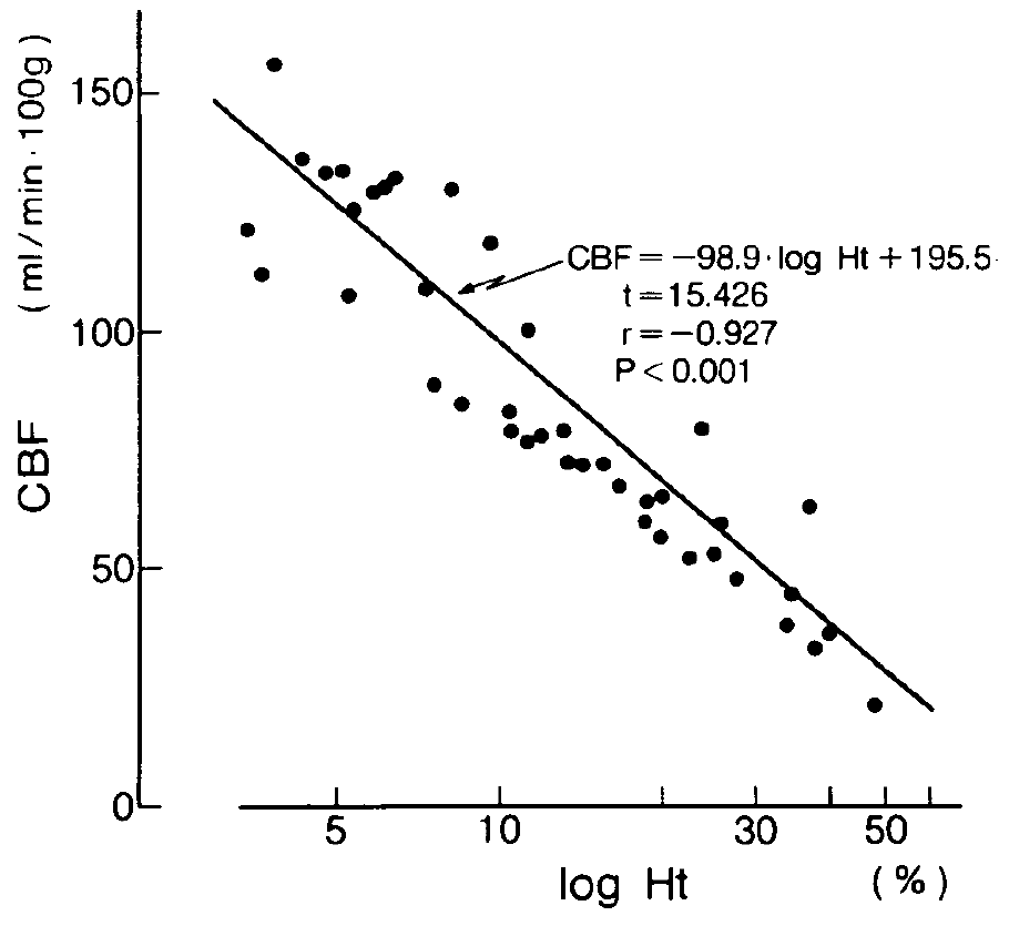
First, we tested the relationship between Ht values and the CBF at a MAP of 100 torr by changing the Ht levels in a wide range (3 to 50%) (normotensive hemodilution) (fig. 1). There was an inverse relationship between Ht and CBF values. A regression line was demonstrated between Ht and CBF values as follows (fig. 1):
CBF (ml/100g·min) = -98.9 log Ht (%) + 195.5
首先,通过大幅改变 Ht 水平(3-50%)(正常血压血液稀释),我们测试了 Ht 值与 MAP 为 100 托的 CBF 之间的关系(图 1)。Ht 和 CBF 值之间有一个反比关系。Ht 和 CBF 值之间有一条回归线,如下所示(图 1):
CBF(ml/100g·min)=-98.9 log Ht(%)+195.5
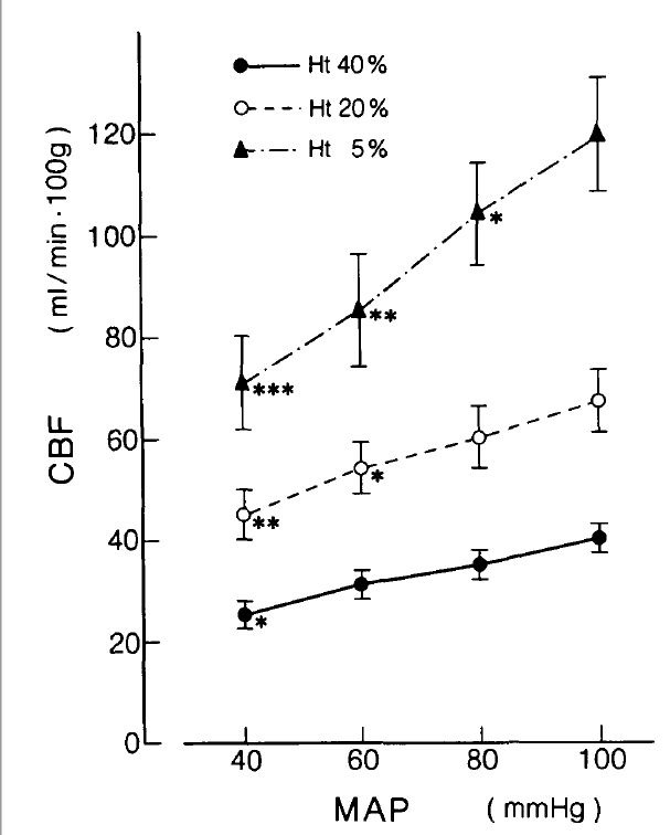
Figure 2 shows a summary of the CBF values at MAPs of 100, 80, 60 and 40 torr during the control (Ht 40), profound (Ht 20) and extreme (Ht 5) hemodilution. At Ht 40, the CBF did not change significantly within the MAP range of 60 to 100 torr, though there was a tendency to decrease as the MAP was lowered. At a MAP of 40 torr, the CBF decreased significantly (p < 0.05). In the profound hemodilution (Ht 20), the CBF increased to about 1.7 times that of the control at a MAP of 100 torr (p < 0.01). As the MAP declined to 60 torr, the CBF decreased significantly (p < 0.05) as compared to that at a MAP of 100 torr in this profoundly hemodiluted state. Thus, the autoregulation of CBF was thought to be already disturbed within this range of the MAP at Ht 20.
图 2 显示了在对照组(Ht 40)、显著(Ht 20)和极端(Ht 5)血液稀释期间,MAP 为 100、80、60 和 40torr 时的 CBF 值的摘要。在 Ht 40 时,CBF 在 MAP 为 60 至 100torr 的范围内没有明显变化,尽管随着 MAP 的降低有下降的趋势。在 MAP 为 40torr 时,CBF 明显下降(P < 0.05)。在显著血液稀释(Ht 20)中,在 MAP 为 100torr 时,CBF 增加到对照组的约 1.7 倍(p < 0.01)。当 MAP 下降到 60torr 时,与 MAP 为 100torr 时相比,在这种显著血液稀释状态下,CBF 明显下降(P < 0.05)。因此,CBF 的自动调节被认为在 Ht 20 时 MAP 的这个范围内已经受到干扰。

FIGURE 1. The relationship between Ht values and the CBF at a MAP of 100 torr in the pentobarbital-anesthetized dogs. The CBF was inversely correlated to the exponent of the Ht values.
图 1. 戊巴比妥麻醉的狗在 MAP 为 100 托时,Ht 值和 CBF 之间的关系。CBF 与 Ht 值的指数呈反比关系。

FIGURE 2. The relationship between the MAP and CBF at 40 (●), 20 (○) and 5% (▲) Ht values in the pentobarbital-anesthetized dogs. CBF values are shown in means and standard errors (bars). The value accompanied by asterisks denotes a significant difference from that at a MAP of 100 torr (* p < 0.05, ** p < 0.01, *** p < 0.001).
图 2. 戊巴比妥麻醉的狗在 40%(●)、20%(○)和 5%(▲)Ht 值时 MAP 和 CBF 之间的关系。CBF 值以平均值和标准误差(条形)显示。伴随星号的数值表示与 MAP 为 100torr 时有明显差异(* p < 0.05,** p < 0.01,*** p < 0.001)。
During extreme hemodilution (Ht 5), the CBF increased to about three times that of control at a MAP of 100 torr (p < 0.001). It decreased significantly at MAPs of 80 (p < 0.05), 60 (p < 0.01) and 40 (p < 0.001) torr in comparison with that at a MAP of 100 torr.
在极度血液稀释期间(Ht 5),CBF 增加到大约是 MAP 为 100torr 时对照组的三倍(p < 0.001)。与 MAP 为 100 托时相比,MAP 为 80(p < 0.05)、60(p < 0.01)和 40(p < 0.001)托时 CBF 明显下降。
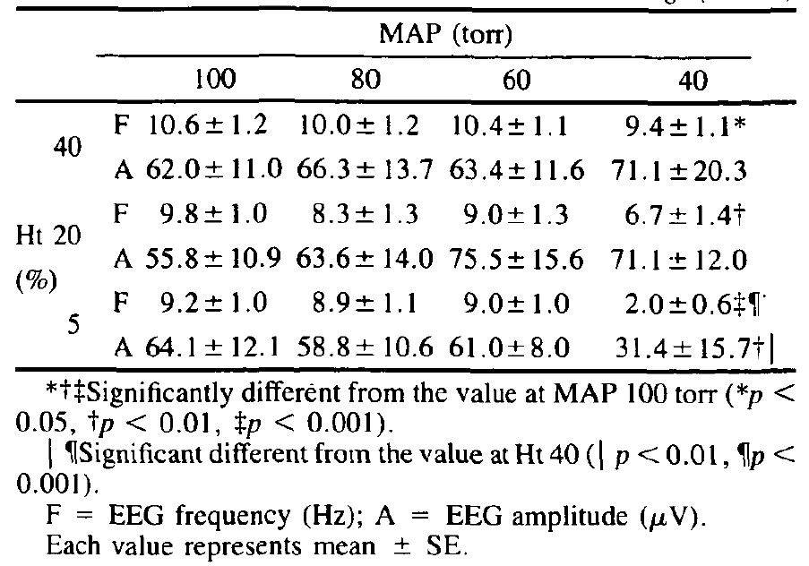
When the MAP was maintained at 100 torr, the changes in the EEG patterns could be barely demonstrated even during extreme hemodilution (Ht 5) (table 1). However, the EEG changes, reflected as slowing of the EEG frequency, became more pronounced during 40 torr of MAP at the HT value of 5% (table 1). Table 1 shows the changes in the EEG frequency and amplitude as a function of the MAP at each Ht value. When the MAP ranged from 100 to 60 torr, the frequency of the EEG did not show any significant change at each Ht value, but revealed a significant slowing during 40 torr of MAP. Further, the EEG amplitude tended to increase as MAP decreased to 40 torr at both Ht 40 and 20 (not significant), while it decreased prominently at Ht 5 under 40 torr of MAP (table 1). Thus, the EEG slowed progressively with reduction in Ht at a MAP of 40 but not at pressures above this level.
当 MAP 维持在 100torr 时,即使在极度血液稀释期间(Ht 5),也几乎看不出 EEG 模式的变化(表 1)。然而,脑电图的变化,反映为脑电图频率的减慢,在 MAP 为 40torr,HT 值为 5% 时变得更加明显(表 1)。表 1 显示了在每个 Ht 值下 EEG 频率和振幅的变化与 MAP 的关系。当 MAP 在 100 到 60torr 之间时,脑电图的频率在每个 Ht 值下没有显示任何明显的变化,但在 MAP 为 40torr 时显示出明显的减缓。此外,在 Ht 40 和 20 时,随着 MAP 下降到 40torr,EEG 振幅有增加的趋势(不显著),而在 MAP 为 40torr 的 Ht 5 时则明显下降(表 1)。因此,在 MAP 为 40 时,脑电图随着 Ht 的降低而逐渐减慢,但在高于这一水平的压力下则没有。

TABLE 1 EEG Frequencies and Amplitudes at Different MAPs with Three Ht Values in Pentobarbital-anesthetized Dogs (n = 8)
表 1 戊巴比妥麻醉的狗在不同 MAP 和三种 Ht 值下的 EEG 频率和振幅(n = 8)。
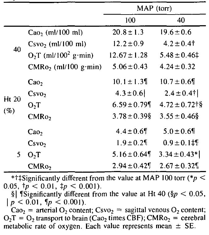
The CMRo2 measured at 100 and 40 torr at each Ht level is shown in table 2, with arterial oxygen content (Cao2), sagittal venous oxygen content (Csvo2) and oxygen transport to brain (O2T). The CMRo2 showed a significant reduction in the hemodiluted states even at a MAP of 100 torr (during isotonic hemodilution). In contrast, there were no significant differences in CMRo2 values between MAPs of 40 and 100 torr in the control as well as in the hemodiluted states. The data in table 2 indicate that the CMRo2 is maintained at MAP of 40 by a considerable increase in oxygen extraction as shown by the reduction in the Csvo,.
表 2 显示了在每个 Ht 水平下 100 和 40torr 测得的 CMRo2,以及动脉含氧量(Cao2)、矢状静脉含氧量(Csvo2)和向大脑输送氧气(O2T)。CMRo2 显示在血液稀释状态下,即使在 MAP 为 100torr 时(等渗血液稀释期间)也有明显的减少。相反,在对照组和血液稀释状态下,MAP 为 40 和 100torr 的 CMRo2 值没有明显差异。表 2 中的数据表明,在 MAP 为 40 时,CMRo2 是通过相当大的氧摄取量来维持的,这表现在 Csvo2 的减少。

TABLE 2 Cao2, Csvo2, O2T and CMRo2 Values at MAPs of 100 and 40 Torr with Three Hi Values in Pentobarbital-anesthesized Dogs (n = 8)
表 2 戊巴比妥麻醉的狗在 MAP 为 100 和 40 Torr 时的 Cao2、Csvo2、O2T 和 CMRo2 值与三个 Hi 值(n = 8)。
# 讨论
The present study has demonstrated that the range of CBF autoregulation becomes narrower as Ht values are reduced. With hemodilution, a progressive increase in CBF has been observed.12-14 This can be caused not only by vasodilatation but also by a blood viscosity reduction.12-14
本研究表明,随着 Ht 值的降低,CBF 自动调节的范围变得更窄。随着血液稀释,已经观察到 CBF 的逐渐增加。12-14 这不仅是由血管扩张引起的,也是由血液粘度降低引起的。12-14
In the presence of extreme hemodilution, the cerebrovascular bed must be submaximally dilated and less responsive to changes in blood gases12 and pressure (incomplete disturbance of the CBF autoregulation). This might account for the differences in the MAPCBF relationship at each Ht value (fig. 2). It has been postulated that the pH of the extracellular fluid of arteriolar smooth muscle is the ultimate mechanism controlling the cerebrovascular caliber.15 According to this hypothesis, cerebral hypoxia caused by hemodilution might reduce the pH of the arteriolar extracellular fluid to produce vasodilatation. This, in turn, might lead to a partial failure of the CBF responsiveness to the changes in MAP as seen in the present study. Besides pH (hydrogen ion), however, the number of candidates for mediating metabolic flow regulation is proposed. At present, adenosine and potassium ion appear to be the most promising other candidates. Adenosine is a strong dilator of pial vessels when applied in the perivascular space.16 Brain adenosine concentration increases under conditions of hypoxia, ischemia, or increased metabolic activity of the brain.17 Similarly, it has been demonstrated that potassium dilates pial arterioles.18
在极度血液稀释的情况下,脑血管床必须是近似扩张的,对血气 12 和压力变化的反应较差(CBF 自动调节的不完全干扰)。这可能是每个 Ht 值下 MAP 与 CBF 关系不同的原因(图 2)。据推测,动脉平滑肌细胞外液的 pH 值是控制脑血管口径的最终机制。15 根据这一假设,血液稀释引起的脑缺氧可能会降低动脉细胞外液的 pH 值以产生血管扩张。反过来,这可能导致 CBF 对 MAP 变化的反应性部分失效,正如本研究中所看到的那样。然而,除了 pH 值(氢离子)外,还提出了介导代谢流量调节的许多候选因素。目前,腺苷和钾离子似乎是最有希望的其他候选因素。在缺氧、缺血或大脑代谢活动增加的情况下,脑腺苷浓度会增加。
The results demonstrated in table 2 reveal that the CMRo2 is very susceptible to changes in Ht values, whereas it is not significantly changed by decreases in the MAP down to 40 torr. On the other hand, the cortical function reflected on the EEG showed no noticeable alterations as a result of profound and extreme hemodilutions when the MAP was maintained at 60-100 torr (table 1), while the EEG slowed in frequency at 40 torr of MAP. The mechanism of this discrepancy between the brain function and the CMRo2 remains to be clarified. A possible explanation may be that the regional distribution of blood flow inside the brain tissue becomes uneven as a result of the decrease in the MAP to 40 torr even when the CMRo2 remains unchanged,4 and this might affect the brain function. Thus, the CMRo2 value did not seem to correlate well with the brain function as detected by electrical activity in hemodiluted states. Deterioration of cerebral activities in terms of the EEG as a result of the decrease in the MAP seemed to correlate more with the rate of the CBF reduction (table 1) than with the CMRo2 values. For instance, EEG frequencies were not significantly different at each Ht value as long as the MAP was maintained at 60-100 torr. This may indicate that the cerebral function can be kept at almost normal levels during normotensive hemodilution at down to 5% of Ht values when MAP is maintained adequately.
表 2 中的结果显示,CMRo2 非常容易受 Ht 值变化的影响,而 MAP 下降到 40torr 时,它没有明显变化。另一方面,当 MAP 维持在 60-100torr 时,反映在 EEG 上的皮质功能没有因显著和极端的血液稀释而出现明显的改变(表 1),而在 MAP 为 40torr 时,EEG 的频率变慢。大脑功能和 CMRo2 之间的这种差异的机制还有待澄清。一个可能的解释是,由于 MAP 下降到 40torr,即使 CMRo2 保持不变,脑组织内血流的区域分布也变得不均匀 4,这可能会影响脑功能。因此,CMRo2 值似乎与血液稀释状态下通过电活动检测的脑功能没有很好的关联。由于 MAP 的降低,脑电活动的恶化似乎与 CBF 降低的速度(表 1)比与 CMRo2 值更相关。例如,只要 MAP 维持在 60-100torr,每个 Ht 值的 EEG 频率就没有明显差异。这可能表明,在正常血压血液稀释期间,当 MAP 充分维持在 5% 的 Ht 值时,大脑功能可以保持在几乎正常的水平。
Recently Fan et al14 have carried out isovolemic hemodilution of up to Ht values of 13% with plasma in dogs in order to measure the responses of alterations in regional hemodynamics and oxygen transport rate. They have demonstrated that oxygen transport to the myocardium does not change significantly at the expense of the increase in coronary blood flow up to an Ht value of 13%, while that to the brain decreases significantly even with an increase in the CBF as the Ht value is reduced to 22%. The MAP, however, was not controlled in their experiment, and systemic arterial pressure was decreased when the Ht value was lower than 20%. The time factor must be also considered in this regard.
最近,Fan 等人 14 用血浆对狗进行了高达 13% Ht 值的等容性血液稀释,以测量区域血液动力学和氧转运率改变的反应。他们证明,在 Ht 值达到 13% 时,以冠状动脉血流增加为代价,心肌的氧转运没有明显变化,而在 Ht 值降低到 22% 时,即使 CBF 增加,大脑的氧转运也明显下降。然而,在他们的实验中没有控制 MAP,当 Ht 值低于 20% 时,全身动脉压下降。在这方面还必须考虑时间因素。
The present experiment has further shown that the CMRo2 is decreased even by normotensive hemodilution without noticeable changes in the EEG when the Ht value is reduced to 20% (table 2). The significant decrease in the CMRo2 without any change in the brain function in terms of EEG might be characteristic in normotensive hemodilution (anemic hypoxia). In contrast, the CMRo2 is reported to be barely affected by a moderate degree of anemia,12 and hypoxic or ischemic hypoxia4 even when the brain function is greatly disturbed. Michenfelder and Theye12 observed no significant change in CMRo2 during hemodilution with plasma expander (dog plasma or low-molecular-weight dextran) down to approximately 15% of Ht value. Discrepancy between their data on CMRo2 and these in the present study might be due to the differences in the solutions infused and/or anesthetics used. In a whole animal, Schwartz et al19 have also suggested that oxygen consumption represents a physiological marker of impending death in the face of progressively diminishing oxygen delivery caused by hypovolemia, anemia, and hypoxia. A more prolonged exposure to hemodilution, however, might have some effect on the EEG in the present experiment. In fact, our previous study3 has shown that reversible slowing of the EEG frequency is noted as a result of extreme normotensive hemodilution (Ht 3-5%) for more than one hour. The present experiment further suggests that an adequate cerebral perfusion is indispensable for the maintenance of the brain function rather than the values of oxygen content in blood or CMRo2.20
本实验进一步表明,当 Ht 值降低到 20% 时,CMRo2 甚至因正常血压的血液稀释而下降,而脑电图没有明显的变化(表 2)。在脑电图方面,CMRo2 的明显下降而脑功能没有任何变化,这可能是正常血压血液稀释(贫血性缺氧)的特点。相反,据报道,CMRo2 几乎不受中等程度的贫血 12 和缺氧或缺血性缺氧 4 的影响,即使大脑功能受到很大干扰。Michenfelder 和 Theye12 观察到在用血浆扩容剂(狗血浆或低分子量葡聚糖)进行血液稀释时,CMRo2 没有明显变化,大约为 Ht 值的 15%。他们关于 CMRo2 的数据与本研究中的数据之间的差异可能是由于输注的溶液和 / 或使用的麻醉剂的不同。在一个完整的动物实验中,Schwartz 等人 19 也提出,在低血容量、贫血和缺氧引起的氧气输送逐渐减少的情况下,耗氧量是即将死亡的生理标志。然而,在本实验中,更长时间的血液稀释暴露可能会对脑电图产生一些影响。事实上,我们以前的研究 3 表明,由于极端的常压血液稀释(Ht 3-5%)超过一小时,脑电图频率会出现可逆的减慢。本实验进一步表明,充分的脑灌注对维持脑功能是必不可少的,而不是血中含氧量或 CMRo2 的数值。
In summary, the present study showed that the range of autoregulation of the CBF became narrower as hemodilution became more profound. Furthermore, the change in the brain function in terms of the EEG seemed to be more correlated with that of the CBF in response to the fall of the MAP than with the CMRo2 value during hemodilution.
总之,本研究表明,随着血液稀释程度的加深,CBF 的自动调节范围变得越来越窄。此外,在血液稀释期间,脑电图方面的变化似乎与 CBF 对 MAP 下降的反应更相关,而不是与 CMRo2 的值相关。
