 关于动脉壁对内部压力变化的局部反应
# 关于动脉壁对内部压力变化的局部反应
ON THE LOCAL REACTIONS OF THE ARTERIAL WALL TO CHANGES OF INTERNAL PRESSURE.
Bayliss WM. On the local reactions of the arterial wall to changes of internal pressure. J Physiol. 1902;28(3):220-231. doi:10.1113/jphysiol.1902.sp00091
关于动脉壁对内部压力变化的局部反应。
Bayliss WM. On the local reactions of the arterial wall to changes of internal pressure. J Physiol. 1902;28(3):220-231. doi:10.1113/jphysiol.1902.sp00091
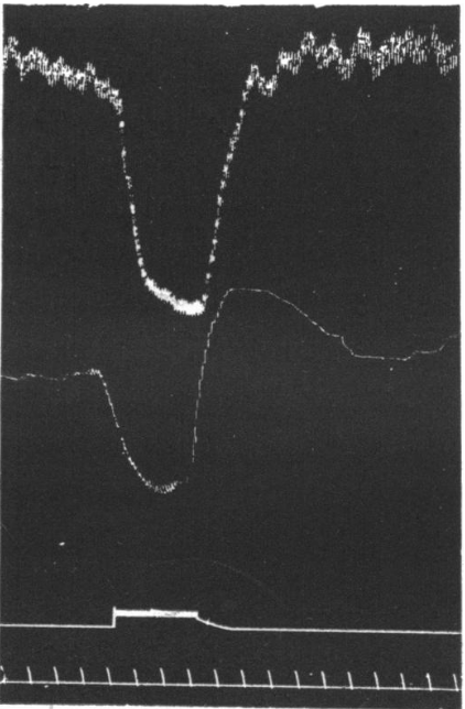
Fig.1.Effect of depressor excitation on volume of enervated leg. Upper curve blood-pressure, next below it volume of leg, upper of two chronographs-period of excitation of depressor nerve, lower one-time in 10 sec.intervals.
图 1. 降压刺激对失去神经支配的腿的体积(粗细)的影响。上面的曲线代表血压,其下为腿的体积,两条时间轴中上面的为 -- 降压神经兴奋期,下面的为 -- 时间间隔为 10 秒一格。
# 序言
MY attention was first directed to these phenomena by the occurrence of curves like that reproduced in Fig. 1
In the course of experiments on vaso-dilator reflexes I sometimes observed what looked like a reflex of this kind, in a limb of which the nerves had been divided; the figure _reproduced shows the effect of a fall of arterial pressure produced by exciting the central end of the depressor nerve on the volume of the hind leg of the rabbit, the sciatic and the nerves accompanying the femoral artery having been cut. It will be noticed that as long as the fall of blood-pressure lasts there is a passive diminution of volume in the limb, but that as soon as the previous height of blood-pressure is attained, on cessation of the excitation, there is a consider-able expansion of the limb lasting for some time. Such a curve would usually be explained by stating that there was present all through the excitation a relaxration of the vessels, but that it was prevented from showing itself in an actual expansion of the limb by the simultaneous fall of blood-pressure; although it was able to produce such an expansion when the blood-pressure rose again owing to the vascular dilatation in other parts, chiefly splanchnic area, lasting a shorter time than in the limb. Such an explanation,obviously, will not hold here, since the leg nerves were cut, and it is plain that some other explanation must be found.
Bearing in mind the well-known reaction of muscle in general to stretching, instances of which are, amongst others, the increased force of the heart-beat produced by increased intraventricular pressure, the effect of tension on the snail's heart, and the contraction of the body walls of the earthworm in response to a pull, it is natural to suppose that the muscular coat of arteries reacts in a similar way to increased intravascular pressure; and if so, being in a state of increased tone in response to the normal blood-pressure, a lowering of that pressure will be followed by an opposite reaction, viz. a relaxation.
This is, no doubt, the explanation of the curve reproduced in Fig. 1. During the fall of blood-pressure the arteries were, in all probability,relaxed in response to that fall, but not to a sufficient extent to produce an actual increase of volume of the limb. This relaxation shows itself at once, however, when the blood-pressure returns to its original level and lasts until the muscular wall of the arteries has responded to the rise of pressure by returning to its original state of tone.
我第一次注意到这些现象是由于出现了类似图 1 的曲线。
在关于血管扩张反射的实验过程中,我有时会在神经被切断的肢体上观察到类似于这种反射的东西;所作的图显示了由压迫神经的中心端产生的动脉压力下降对兔子后腿体积的影响,坐骨神经和伴随股动脉的神经被切断。我们会注意到,只要血压下降并维持低血压,肢体的体积就会逐渐缩小,但一旦达到之前的压力水平,停止刺激后,肢体就会出现相当程度的膨胀,并持续一段时间。这样的曲线通常被这样解释:在整个兴奋过程中存在血管舒张,但由于血压同时下降,这种舒张没能发展为肢体的实际膨胀。尽管由于其他部位的血管舒张,主要是内脏,当血压回升时,相应部位能够表现出膨胀,持续时间要比四肢短。显然,这种解释在这里不成立,因为腿部神经被切断了,很显然必须找到其他解释。
考虑到众所周知的肌肉对拉伸的通常反应,其中的例子包括:因心室内压力升高而产生的心跳力量增加,压力对蜗牛心脏的影响,以及蚯蚓的体壁因受拉而收缩,我们很自然地认为,动脉的肌层对血管内压力升高也有类似的反应。如果是这样,由于对正常血压所作出的反应,血管处于压力升高的状态,那么压力降低后就会出现相反的反应,即舒张。
毫无疑问,这就是对图 1 所呈现曲线的解释。在血压下降的过程中,动脉很可能因血压下降而舒张,但舒张的程度不足以产生肢体容积的实际增加。然而,当血压恢复到原来的水平时,这种舒张立即表现出来,并持续到动脉的肌肉壁对压力上升的反应,恢复到原来的压力状态。
# 对压力升高的反应
Since this reaction is the more familiar one in other muscular tissues, I will first describe some experiments made to detect it in arterial muscle. So far as I am aware, indeed, the opposite form of reaction, viz. relaxation to diminished tension, has not, hitherto, been described. It must obviously occur, however, in a muscle in a state of tone, if that tone is present in response to a condition of tension, when the exciting cause of the tone, viz. the tension, is diminished.
To produce a rise of arterial pressure in these experiments, in order to provoke the response, one can either excite the peripheral ends of the splanchnic nerves, or produce a reflex rise by exciting the central end of a pressor sensory nerve, or by producing asphyxia in a curarized animal. In the two latter cases, naturally, the vaso-motor nerves of the organ must be cut, as also in the case of splanchnic excitation, when a viscus supplied by that nerve is under observation. As regards the limbs I have not noticed that there is any difference in the reaction whether the nerves are cut or not. The reaction is, therefore, of peripheral origin, and, as I believe, myogenic in nature; but this point will be discussed later.
The animals used, dogs, cats, and rabbits, were anesthetized with morphia (30 to 130 mgrms. subcutaneously) and kept under A.C.E. In most cases curare was also given.
由于这种反应在其他肌肉组织中是比较熟悉的,我将首先描述在动脉肌中检测它的一些实验。据我所知,与此相反的反应形式,即在压力下降时的舒张,迄今为止还没有被描述。然而,对于处于紧张状态的肌肉,如果这种紧张是对紧张状态的反应,当引起紧张的原因(即紧张)消失时,显然一定会出现舒张。
为了在这些实验中产生动脉压力的上升,以激起反应,我们可以刺激内脏神经的外周端,或者通过刺激升压感觉神经末端中心,或者令箭毒瘫痪的动物出现窒息来产生反射性上升。在后两种情况下,自然必须切断该器官的血管运动神经。当观察的内脏组织的神经支配被用作内脏兴奋刺激时,也要切断。至于四肢,我没有注意到神经被切断与否,在反应上有什么不同。如此一来,观察的反应只能是是起源于外周的,而且,我认为本质是肌源性的;稍后讨论这点。
所用的动物,包括犬、猫和兔子,都是用吗啡麻醉的(皮下注射 30 至 130 毫克),并在 A.C.E. 下保存。多数情况也给予箭毒。
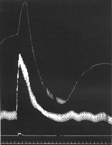
Fig. 2. Effect of splanchnic excitation on enervated limb of dog. Upper curve-volume of limb. Lower curve-blood-pressure, zero being 45 mm. below excitation marker, which is the upper of the two bottom lines. Time in 10".
图 2. 内脏刺激对犬去神经支配的肢体的影响。上部曲线为 -- 肢体体积。下部曲线代表 -- 血压,横坐标零点血压为 45 毫米。下方为刺激标志,是两条底线的上面那条。时间为 10 秒一格。
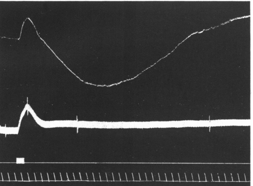
Fig. 3. Effect of splanchnic excitation on the enervated hind-leg of cat. Explanation of Fig. 2 applies also to this.
图 3. 内脏刺激对猫失去神经支配后腿的影响。
图解同图 2。
The curves of Fig. 2 show the effect of exciting the peripheral end of the cut splanchnic nerve on the volume of the enervated hind-leg of the dog. As the arterial pressure rises the limb is distended passively,but instead of merely returning to its original volume when the blood-pressure has come down again, it constricts much below its previous level and only gradually returns.
Fig. 3 shows a similar effect in the cat.
In asphyxia one occasionally meets with curves like Fig. 4. In this case the leg nerves were cut, and at the point A the artificial respiration was stopped. At first the leg is passively distended, but, while the blood-pressure is still at its height, the limb begins to diminish in volume, attaining its original size while the blood-pressure is consider-ably above its previous height. Of course the effect in this case is possibly complicated by the admixture of the direct action of asphyxial blood on the vessels, but there is reason to believe that the latter is rather of the nature of a dilatation than constriction, although the point is still uncertain.
图 2 的曲线显示了刺激被切断的内神经外周端对犬的后腿的体积的影响。当动脉压力上升时,肢体被动地膨胀,但当血压再次下降时,它不是仅仅恢复到原来的体积,而是缩小到低于以前的水平,并逐渐恢复。
图 3 显示了猫的一个类似效果。
在窒息情况下,人们偶尔会遇到像图 4 这样的曲线。在这种情况下,腿部神经被切断,并在 A 点停止人工呼吸。起初,腿部是被动膨胀的,但是,当血压仍较高时,肢体开始缩小,达到原来的大小,而血压却大大高于其以前的高度。当然,这种情况下的效果可能因为混合了窒息性血液对血管的直接作用而变得复杂,但有理由相信,后者的性质是扩张而不是收缩,尽管这一点仍不确定。
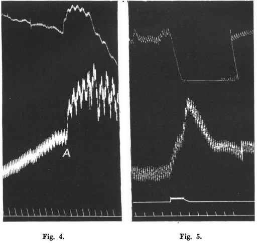
Fig. 4. Effect of asphyxial rise of blood-pressure on volume of enervated leg. Same explanation as previous figures. Blood-pressure zero-13 mm. below time tracing. At A artificial respiration stopped, the animal being under curare.
图 4. 窒息导致血压上升对神经离断腿的体积的影响。图解如前。零点血压为 --13 mm,下面是时间线。A 处人为中断呼吸,动物经箭毒化处理。
Fig. 5. Effect of splanchnic excitation on volume of kidney of dog. Upper curve-volume of kidney. Lower curve-blood-pressure. Excitation and time markers as previous figures. Blood-pressure zero-55 mm. below excitation marker.
图 5. 内脏刺激对犬的肾脏体积的影响。上部曲线:肾脏体积。下部曲线:血压。刺激和时间标记与前图相同。零点血压:55mm,下面为刺激标记。
In the case of the kidney vessels this reaction to increased tension is very marked, as shown in Fig. 5. All the nerves in the hilus that could be found were torn through, the vessels and ureter thoroughly painted with concentrated phenol, which I have found a very useful means of destroying the conductivity of nerve-fibres. The vessels and ureter, moreover, were tightly pinched with forceps, at the risk of causing the blood to clot, which fortunately, however, did not take place until the experiment had been performed. The kidney was enclosed in a Roy's oncometer, filled with warm physiological saline instead of oil, the fluid only reaching to the beginning of the tube leading to the piston-recorder, which contained air. The splanchnic nerve of the same side was exposed in the abdomlinal cavity and on Ludwig electrodes. The rise of blood-pressure produced by exciting the nerve is seen to cause at first a slight passive distension of the kidney, but even when the pressure is still rising the kidney begins to react by constriction which carries the piston of the recorder down to the limit of its excursion. The drum was stopped for a short time to allow the lever to return to its original position. The only source of fallacy that I can see in these experiments is the possible escape of current from the electrodes on the splanchnic nerve to the kidney vessels directly. In order to exclude this I made another experiment in which the lower end of the divided spinal cord was excited, the cord was cut at the level of the 4th thoracic nerve, and the lower end excited in the spinal canal (Fig. 6).
The kidney nerves had been cut,painted with phenol and tightly pinched with forceps. The reaction is not so striking as in Fig. 5, but there is an obvious constriction of the organ,while the blood-pressure was at its height. The circulation through the kidney was not so good in this case as in that of the previous figure,since the vessels had been pinched a longer time before the experiment was made, and had probably become partially obstructed with clot.
如图 5 所示,在肾脏血管,这种对压力升高的反应非常明显。所有可以找到的肾门中的神经都被完全撕开,血管和输尿管被彻底涂上了浓缩的苯酚,我发现这是一种非常有用的破坏神经纤维传导性的手段。此外,还用镊子紧紧夹住血管和输尿管,有血液凝固的风险,然而幸运的是,在实验进行之前,并没有发生凝血。肾脏被装在罗氏器官体积测量器中,里面装的是温热的生理盐水而不是油,液体平面刚好到一个管子开口处,管子连接活塞记录器,里面有空气。肾脏同侧的内脏神经暴露在腹腔内和路德维希电极上。通过刺激神经而产生的血压上升,可以看到一开始会引起肾脏的轻微被动膨胀,但即使在压力仍在上升,肾脏已开始作出收缩反应,从而使记录器的活塞下降到其行程的极限。鼓停了一小会儿,让杠杆回到原来的位置。在这些实验中,我所看到的唯一缺陷是电流可能从内脏神经上的电极直接逃到肾脏血管。为了排除这一点,我又做了一个实验,在这个实验中,刺激分离的脊髓下端,脊髓在第四胸神经水平被切断,刺激椎管内的脊髓下端(图 6)。
肾脏神经已被切断,用苯酚涂抹,并用镊子紧紧夹住。反应没有图 5 那么明显,但器官有明显的收缩,而血压处于最高水平。在这种情况下,通过肾脏的血液循环并不像上一个图中的那么好,因为在进行实验之前,血管已经被挤压了很长时间,而且可能已经被血块部分阻塞了。
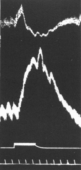
Fig. 6. Effect of exciting spinal cord on volume of enervated kidney. Explanation as previous figures. Blood-pressure zero 23 mm. below excitation marker.
图 6. 刺激脊髓对去神经支配肾脏体积的影响。图解如前。横坐标零点的血压为 23mm。下面是刺激标志。
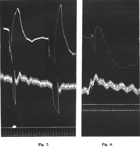
Fig. 7. Effect of compression of the abdominal aorta on the volume of the hind-limb. Explanation of curves as before. Blood-pressure zero at level of excitation marker. Two compressions, the second not marked by the signal.
图 7. 压迫腹主动脉对后肢体积的影响。图解同前。血压零点位于刺激标记水平处。两次按压,第二次没有用符号标记出来。
Fig. 8. A similar curve to Fig. 7. Blood-pressure cannula in this case in carotid. Leg nerves cut. At x, obstruction of abdominal aorta. Blood-pressure zero-48 mm. below excitation marker.
图 8. 与图 7 的曲线类似。本例中的血压导管位于颈动脉。腿部神经被切断。符号 X 处代表阻断腹主动脉。零点血压:48mm,下面是激发标记
# 对压力下降的反应
We may now consider the opposite form of reaction, viz. the relaxation produced by diminution of blood-pressure.
Fig. 7 is an example of the effect produced on the volume of the hind-limb by aoitic obstruction. The cannula recording the arterial pressure was in the femoral artery of the opposite side, so that its excursions record the actual pressure in the artery of the limb under observation. This dog bad been used for experiments on vascular reflexes, so that the sciatic and anterior crural nerves were not cut, but it was cut off from the vaso-motor centre by extirpation of the abdominal sympathetics on both sides and by section of the cord at the 2nd lumbar vertebra. There is seen to be a large dilatation of the limb, following the rapid diminution of volume, produced by compression of the abdominal aorta by means of a thread on a ligature-staff. There is also seen here a constriction following the dilatation. This is still better shown in Fig. 8, and is no doubt to be explained by the sudden inrush of blood at high pressure into the relaxed arterioles causing a constrictor reaction, of the kind described in the earlier part of this paper, so that there is, so to speak, a kind of reverberation. This second reaction is also well shown in Fig. 9., an example from the cat.
我们现在可以考虑相反的反应形式,即由血压降低产生的舒张。
图 7 是一个例子,说明腹主动脉阻塞对后肢容积产生的影响。记录动脉压力的插管在对侧的股动脉中,因此它的偏移记录了被观察肢体动脉中的实际压力。这只犬曾被用于血管反射的实验,因此坐骨神经和前胸神经没有被切断,但它通过切除两侧的腹部交感神经和在第二腰椎处切除脊髓而与血管运动中心切断。我们可以看到,通过结扎器上的线扎紧腹主动脉,肢体体积迅速缩小,随后膨胀明显变大。这里还可以看到扩张后的收缩。这一点在图 8 中显示得更好,毫无疑问,这可以解释为血液在高压下突然涌入舒张的动脉血管,引起收缩反应,就像本文前面所描述的那样,因此可以说有一种回响震荡。这第二种反应在图 9 中也有很好的显示,这是一个来自猫的例子。
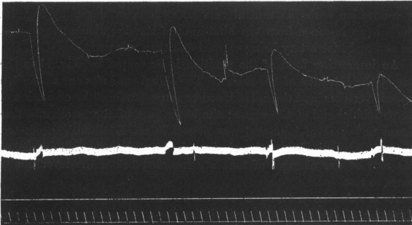
Fig. 9. Four cases of compression of aorta producing double reaction in the enervated hind-limb of t-he cat. Blood-pressure zero-45 mm. below excitation marker.
图 9. 四次压迫主动脉在猫的后肢上产生的双向反应。零点血压为:45 mm,下面为激发标记。
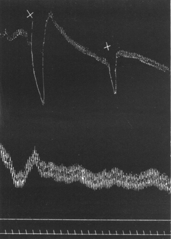
Fig. 10. Effect of compression of iliac artery on volume of limb with nerves intact. Explanation as previous figures. Artery clipped twice at x. Blood-pressure zero-50 mm. below excitation marker.
图 10. 压迫髂动脉对神经支配完好的肢体体积的影响。图解同前。在符号 X 处动脉被压迫两次,零点血压为:50 mm,下面为激发标记。
As remarked previously the reaction in question is not prevented by connection with the central nervous system, as shown by Fig. 10, which is an instance from the hind-limb of the dog with nerves intact, the external iliac artery being clipped and then released.
An interesting form of the reaction is presented by Fig. 11 in the case of the intestine of the rabbit. The spinal cord was cut at the 4th thoracic vertebra, and the fall of blood-pressure produced by excitation of the peripheral end of the vagus; the lower part of the small intestine (ileum and part of jejunum) was placed in an Edmunds' oncometer. The " reverberation " mentioned above is shown in a marked degree, so that four or five changes of tone are recorded before the lever finally comes to rest.
如前所述,有关反应并不因与中枢神经系统的联系而被阻止,如图 10 所示,这是一个来自神经完好的犬的后肢的例子,髂外动脉被压迫,然后释放。
图 11 是一个有趣的反应形式,是在兔子的肠子上看到的。在第四胸椎处切断脊髓,通过激发迷走神经的外周端产生血压下降;将小肠的下部(回肠和空肠的一部分)放在 Edmunds 的测压器中。上面提到的 "混响" 显示出明显的程度,因此,在杠杆最终静止之前,会记录下四或五次的音调变化。
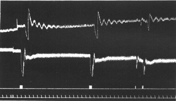
Fig. 11. Effect of excitation of peripheral end of the vagus on the volume of the intestine. Upper curve-volume of intestine. Lower curve-blood-pressure. Excitation and time-markers as previous figures. Blood-pressure zero, at level of excitation marker.
图 11. 迷走神经末梢的兴奋对肠道容积的影响。上部曲线:肠道容积。下部曲线:血压。刺激和时间标记同前图。血压零点处于刺激标记的水平
As a rule the reaction is not shown in a convincing manner by excitation of the peripheral end of the vagus, since the fall of blood- pressure so produced is usually followed by a rise, due partly to increased action of the heart, partly to slight asphyxial excitation of the vaso-motor centre, and this rise may be thought to be the cause of the expansion of the organ. In Fig. 11, however, it will be noticed that the rise of blood-pressure does not occur in the second excitation of the vagus, although the dilator reaction of the intestinal vessels is very marked.
It is necessary to meet a possible objection. It may be thought that the relaxation described is due to anaemia causing asphyxia of the arterial wall. That this is not so is shown by the fact that I have repeatedly seen it in a curarized animal, in which, owing to excessive artificial respiration, there was no rise of blood-pressure on stopping the pump until more than a minute had elapsed. The tissues were there-fore copiously supplied with oxygen, and the obstruction of the artery produced the reaction if lasting only for a couple of seconds. In the case of the limb, moreover, it must be remembered that the muscle metabolism in a curarized animal is very small, and that a prolonged anaemia would be necessary to cause an appreciable degree of asphyxia. This objection is also answered by the experiments on excised arteries to be described below.
通常情况下,迷走神经末梢的兴奋并不能令人信服地显示出这种反应,因为这样产生的血压下降通常会随之上升,部分是由于心脏的作用增强,部分是由于血管运动中心的轻微窒息性兴奋,这种上升可能被认为是器官扩张的原因。然而,在图 11 中,我们可以注意到,在迷走神经的第二次兴奋中并没有出现血压的上升,尽管肠道血管的扩张反应非常明显。
有必要回答一个可能的反对意见。可能有人认为,所述的舒张是由于贫血导致动脉壁窒息。事实证明并非如此,我曾多次在一个箭毒化处置的动物身上看到这种情况,由于过度的人工呼吸,在停止泵的时候,血压没有上升,直到超过一分钟后。因此,组织得到了大量的氧气供应,而动脉的阻塞产生了反应,尽管只持续了几秒钟。此外,对于肢体,必须留意,箭毒化处置的动物的肌肉代谢是非常小的,长时间的贫血才能导致明显程度的窒息。下面将介绍的切除动脉的实验也回答了这一异议。
# 在切除的动脉中的反应
It has been already pointed out above that these reactions are independent of the central nervous system, and are therefore of peripheral origin. Since no ganglia have been found in connection with the blood vessels it follows that the effects in question are of myogenic nature, as indeed one would expect from the known properties of smooth muscle. But, in view of Mac William's researches on excised arteries, it seemed worth while to attempt to obtain re- actions on them. Owing to the pressure of other work I have not been able to make many experiments of this kind. I have, however, obtained reactions, and in one case the effect was so marked as to be visible without the aid of any instrumental magnification. This case was that of a carotid artery removed from the body of a dog three hours after death by asphyxia, so that, if there were any ganglion-cells attached to the artery, they were presumably quite out of action. A cannula was inserted in one end of the artery, and the other end tied; the artery and cannula were filled with defibrinated blood and connected by india-rubber tubing to a mercury reservoir which could be raised and lowered. When now the pressure was raised inside the artery it was seen at first to swell, but immediately, and while the mercury was still kept at its height, a powerful contraction took place, in which the artery appeared to writhe like a worm. If the pressure was suddenly lowered again the artery did not return merely to the state corresponding to the lesser pressure, but underwent a considerable relaxation, which passed off just as in the vessels of the body as a whole. This reaction to lowered pressure was better seen when the artery was inclosed in a small air oncometer, consisting of a wide glass tube connected with a piston recorder, by means of which an enlarged curve of the changes of volume could be obtained. I regret that the tracing was spoilt in varnishing, so that I am unable to reproduce it here.
The fact of the occurrence of the reaction in an excised and asphyxiated artery is of importance for three reasons:--
- It disposes of the objection that the phenomena in question are due to any other cause than the change of tension.
- It affords a proof of their myogenic nature.
- In the particular experiment described above it shows that the contraction in response to increased pressure may show itself while the pressure continues, so that the form of curve given by the kidney,which I for some time looked upon with distrust, is quite a possible one, even in the complete absence of any excitation of vaso-constrictor nerve-fibres.
上面已经指出,这些反应是独立于中枢神经系统的,因此是外周性的。由于没有发现与血管有关的神经节,因此有关的影响是肌源性的,正如人们从平滑肌的已知特性中所期望的那样。但是,鉴于麦克 - 威廉对切除的动脉所做的研究,似乎值得尝试在这些动脉上获得再作用。由于其他工作的压力,我没能做很多这样的实验。然而,我已经获得了反应,在一个案例中,效果非常明显,不需要任何仪器的放大就可以看到。这个案例是在一只犬因窒息死亡三小时后从其体内取出的颈动脉,因此,如果有任何神经节细胞附着在动脉上,它们大概已经完全失去了作用。在动脉的一端插入插管,另一端绑住;动脉和插管中充满了去纤维蛋白的血液(防止凝固),并通过印度橡胶管连接到一个可以升高和降低的水银计。此时当动脉内的压力升高时,起初看到的是肿胀,但是当水银仍保持在其原有高度时,动脉立即发生了强有力的收缩,过程中动脉似乎像蠕虫一样蠕动着。如果压力突然再次降低,动脉并不只是恢复到与较低压力相对应的状态,而是出现了相当大程度的舒张,随后这种舒张消失了,就像在整个身体的血管一样。当动脉被置于一个小型的空气器官体积测量仪中时,可以更好地看到这种对压力降低的反应,该测量仪由一根宽大的玻璃管和一个活塞记录器组成,通过它可以获得体积变化的放大曲线。遗憾的是,描记图在上漆时被破坏了,所以我无法在此复制它。
在离体动脉和窒息下动脉实际发生的反应是很重要的,原因有三:
- 它平息了反对意见,反对意见认为有关现象是由压力变化以外的任何其他原因造成的。
- 它为其肌源性提供了一个证据。
- 在上面描述的特殊实验中,它表明在压力持续的情况下,压力升高所对应的收缩现象在高压继续保持时可能会显示出来,因此,由肾脏给出的曲线形式,我有一段时间不相信,是相当可能的,甚至出现在完全没有任何血管收缩神经纤维的兴奋下。
# 反应的意义
It remains to indicate briefly how the behaviour of the blood vessels described in the preceding article may be of service in the normal organism.
The peripheral powers of reaction possessed by the arteries is of such a nature as to provide as far as possible for the maintenance of a constant flow of blood through the tissues supplied by them, whatever may be the height of the general blood-pressure, except in so far as they are directly overruled by impulses from the central nervous system.
As examples let us take first the case in which, say for cardiac reasons, a considerable general fall of blood-pressure is required. This,as we know, is produced by universal vascular dilatation (except in the brain); but, owing to the preponderance of the splanchnic area, the limbs, the relaxation of whose vessels is usually insufficient to produce an actual increase of their sectional area under the low arterial pressure prevailing, would be deprived of their due supply of blood when perhaps it might be particularly needed, were it not that their arterioles automatically relax to a still greater degree, and so make up for the lowered pressure by an increase of sectional area.
Take again the case in which as great as possible a rise, or fall, of arterial pressure is required; if the limb-vessels allowed themselves to be passively distended, or drained, as the case may be, the result would be to partially annul the effect of constriction, or dilatation, in the splanchnic area, but, since they react in the way described, they assist in the production of the desired end.
An especially interesting case is that of the cerebral circulation. There is, up to the present, no satisfactory evidence of the presence of a vaso-motor supply to the brain; so that when an increased blood-supply is required in that organ it is provided by constriction of vessels throughout the rest of the body, which is, in this respect, so to speak the slave of the brain. Now Hill has shown that the tendency of increased arterial distension in the brain is to produce cerebral anaemia, owing to the skull being a rigid box, so that, although the arteries may increase in size, it is at the expense of the capillaries and veins;. this swelling of the arteries is prevented by the automatic reaction of their muscular coat, as described above, so that they do not allow themselves to be distended, but become more or less rigid, and so permit the increased velocity of flow, due to increased pressure, to produce its desired effect in providing a more abundant blood-supply to the active cerebral tissue.
On the other hand it is evident that the effects of a rise of general blood-pressure of a moderate degree, produced, say, by constriction of the splanchnic area, would be to cause automatic contraction of the arteries of all other parts of the body, and thus raise still further the general blood-pressure, unless dilator impulses were sent to the vessels from the central nervous system. Thus, without the controlling government of the latter, every rise of pressure would automatically cause a further rise, and every fall, from whatever cause, a further fall of blood-pressure from peripheral vascular dilatation; i.e. a vicious circle would be established in the absence of the regulating functions of the vaso-motor centres.
我们仍需简要说明前文所述的血管行为在正常机体中是如何发挥作用的。
动脉所拥有的外周反应能力的性质是,无论总体血压多高,都能尽可能地维持血液持续流经由它们供应的组织,除非它们直接被来自中枢神经系统的冲动所支配。
作为例子,让我们先看看这样的情况,比如说由于心脏的原因,前提是总体血压下降非常明显。我们知道,这是由普遍的血管扩张产生的(脑部除外);但是,由于内脏区域的优势,四肢的血管舒张通常不足以在那种动脉低压水平下产生实际横截面积的增加,如果不是它们的动脉血管自动舒张到更大的程度,从而通过面积的增加抵消血压下降引起的血流减少,在全身特别需要的时候,它们可能会被剥夺应有的血液供应。
再以需要尽可能大的动脉压力上升或下降的情况为例;如果肢体血管允许自己被动地膨胀或排空,视情况而定,其结果将是部分地取消收缩或扩张的影响,但是,在内脏区域,由于它们以所述方式作出反应,它们有助于产生所需的结果。
一个特别有趣的例子是脑循环的例子。到目前为止,还没有令人满意的证据表明大脑存在血管运动支配;因此,当该器官需要增加血液供应时,它是通过收缩整个身体其他部位的血管来提供的,在这方面,可以说全身血管是大脑的奴隶。现在,希尔已经证明,由于头骨是一个坚硬的盒子,因此,尽管动脉可能会增大,但它是以牺牲毛细血管和静脉为代价的,因此,大脑中动脉扩张的趋势是产生脑缺血;如上所述,动脉的这种肿胀被其外被肌肉的自动反应所阻止,因此它们不允许自己膨胀,而是或多或少地变得僵硬,因此允许由于压力升高而导致的流速增加,从而产生预期效果,为活跃的大脑组织提供更丰富的血液供应。
另一方面,已证明,适度的总体血压上升,将导致身体所有其他部位的动脉自动收缩,(例如由内脏区域的收缩产生的影响),从而进一步提高总体血压。除非中枢神经系统向血管发出舒张冲动。如果没有神经控制,每一次压力的上升都会自动导致血压进一步的上升,而每一次下降,不管是什么原因,都会导致外周血管扩张导致血压进一步下降;也就是说,如果没有血管运动中心的调节功能,就会形成一个恶性循环。
# 总结
The muscular coat of the arteries reacts, like smooth muscle in other situations, to a stretching force by contraction.
It also reacts to a diminution of tension by relaxation, shown, of course, only when in a state of tone.
These reactions are independent of the central nervous system, and are of myogenic nature.
They are to be obtained both in the case of vessels in their normal condition in the body, and in excised arteries some hours after death.
- 与其他情况下的平滑肌一样,包裹动脉的肌层通过收缩来对抗牵张力。
- 压力下降时动脉的反应是舒张,当然只在动脉有张力时。
- 这些反应独立于中枢神经系统,是肌源性的。
- 在体内正常状态下的血管和死后几小时内切下的动脉中,都可以得到这些反应。
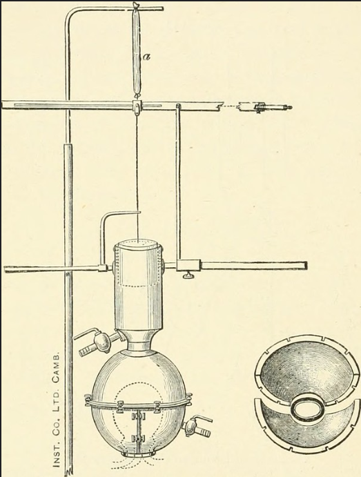
Image from page 265 of "Elements of human physiology" (1907)
Diagram of Roy's cardiometer. On the right of the figure are the two quarter spheres which are clamped on to the pericardium at the root of the heart. itself filled with oil or air as the oncometer, and register the changes in the volume of the heart by connecting the cavity of the pericardium with some form of piston-recorder, or we may make use of Roy's cardiometer. This is a brass sphere in three segments. The two quarter spheres (Fig. 142) are applied round the base of the heart and clamped together, the cut pericardium being attached to their constricted neck, the third segment, a hemisphere, is then applied over the
